Manuel Dias de Abreu: The Brazilian Doctor Who Revolutionized Tuberculosis Screening
When we think about medical pioneers, we often remember the scientists who discovered a disease, invented a medicine, or developed a life-saving surgical procedure. But some of the most important contributions to medicine come from people who find better ways to detect disease before it becomes fatal.
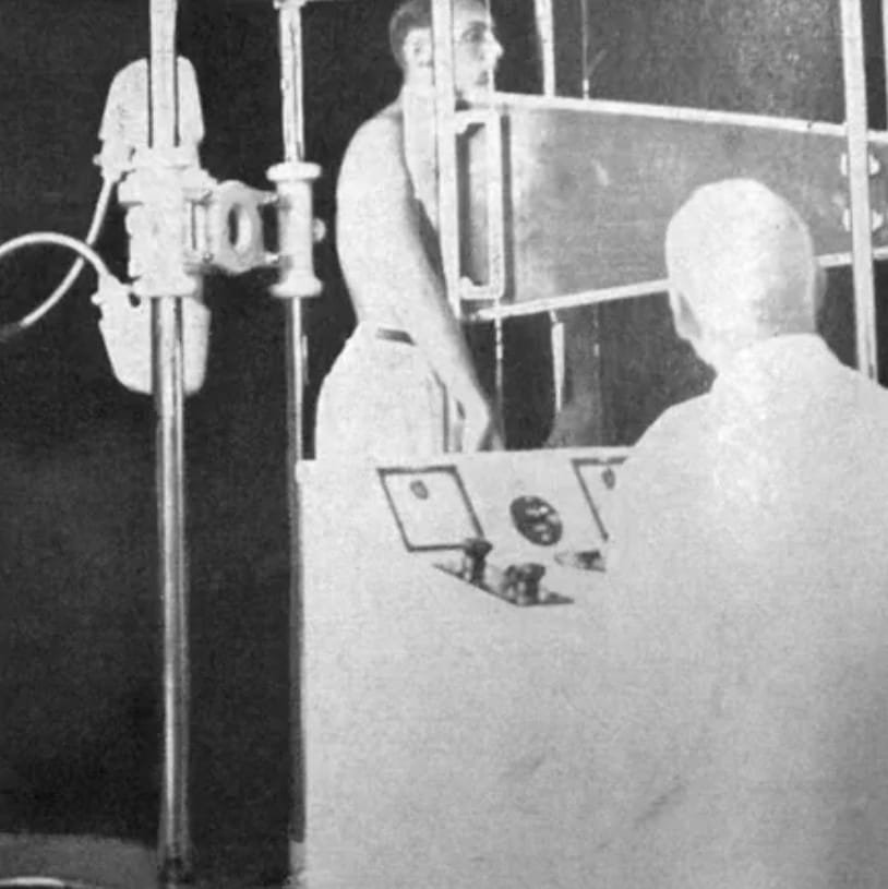
One such pioneer was Manuel Dias de Abreu, a Brazilian physician, radiologist, scientist, and inventor whose work transformed the mass detection of tuberculosis. His most famous creation, abreugraphy, was a miniature chest-radiography technique designed to make large-scale screening faster, more practical, and less expensive.
At a time when tuberculosis was a major public-health threat, Abreu recognized that early diagnosis could make an enormous difference. His invention eventually became an important tool in tuberculosis-control programs in Brazil and other countries. The importance of his work was also recognized internationally: the Nobel Prize archive records six nominations for Manuel de Abreu, including four in 1946, one in 1951, and one in 1953.
Early Life and Education
Manuel Dias de Abreu was born in São Paulo, Brazil, on January 4, 1891, according to the Brazilian National Academy of Medicine. His parents were Júlio Antunes de Abreu, who was Portuguese, and Mercedes da Rocha Dias, from São Paulo.
Abreu studied medicine at the Faculty of Medicine of Rio de Janeiro. He completed his medical degree in 1914, presenting a thesis titled Natureza pobre. From an early stage in his career, however, he was particularly interested in radiology.
Radiology was still a developing field during Abreu’s youth. X-rays had opened an entirely new window into the human body, allowing doctors to see structures that previously could only be examined indirectly. Abreu understood that this technology could become particularly valuable in diseases affecting the lungs.
After completing his medical studies, Abreu traveled to Europe with his family. He spent time in Lisbon and then in Paris, where he developed his expertise in radiology. He became associated with major medical institutions, including the Hôtel-Dieu and Hospital Laennec. At Hospital Laennec, he concentrated on pulmonary radiology, studying the differences in tones and shadows visible in chest images.
His European experience gave him a strong scientific foundation. More importantly, it prepared him for the medical problem that would define his career.
Returning to Brazil
In 1922, Abreu returned to Rio de Janeiro. What he encountered deeply influenced his future work.
Tuberculosis was causing enormous suffering in the city. The disease could progress silently, and many patients sought medical attention only after it had reached an advanced stage. By then, treatment was much more difficult.
Abreu became convinced that one of the keys to controlling tuberculosis was early detection.
He insisted on developing better radiological services for tuberculosis patients. Eventually, the first radiology service in Rio de Janeiro specifically dedicated to the disease was created, and Abreu headed the X-ray Department of the Tuberculosis Prophylaxis Inspectorate.
The challenge was not simply taking an X-ray of one patient. Abreu wanted a technique that could potentially be used to examine large numbers of people.
That was a much bigger technological problem.

The Tuberculosis Challenge
Tuberculosis is caused by Mycobacterium tuberculosis and primarily affects the lungs. Before modern antibiotics became widely available, tuberculosis was one of the world’s most feared infectious diseases.
Doctors needed ways to identify infected people before severe symptoms appeared. Chest radiography offered an important possibility because tuberculosis could produce visible abnormalities in the lungs.
But conventional radiography had limitations when used for mass screening. Equipment was expensive, examinations required time, and producing full-size radiographs for enormous populations was not always practical.
Abreu began looking for a solution.
His goal was simple in principle:
Could doctors obtain useful chest images quickly enough to screen thousands of people?
The answer would become one of his greatest achievements.
The Birth of Abreugraphy
Abreu developed a technique that became known as abreugraphy.
The method was different from conventional radiography. Instead of producing a traditional full-size radiograph, the technique involved photographing the image produced during fluoroscopic examination, creating a small image that could be rapidly produced and examined.
The Brazilian National Academy of Medicine describes the technique as a form of indirect chest radiography and notes that Abreu presented his invention to the Society of Medicine and Surgery of Rio de Janeiro in 1934.
This was an important development because the smaller images could make mass screening considerably more practical.
The technique was initially associated with tuberculosis detection, but its usefulness eventually extended beyond tuberculosis. According to the Brazilian National Academy of Medicine, abreugraphy was also used in identifying lesions involving the heart, major blood vessels, and lung tumors.
Why the Invention Was Important
The brilliance of Abreu’s work was not simply that he created another type of X-ray image.
His achievement was connected to the idea of population-level medicine.
Traditional medical diagnosis often focuses on an individual patient who visits a doctor because of symptoms. Abreu’s approach helped move radiology toward another model: examining large groups of apparently healthy people to identify disease at an earlier stage.
This was particularly valuable for tuberculosis.
If a person with early pulmonary tuberculosis could be identified through screening, that person could potentially receive medical attention sooner. At the same time, identifying cases earlier could help public-health authorities respond to a contagious disease.
Abreugraphy therefore represented a meeting point between radiology, preventive medicine, and public health.
Recognition of the Name “Abreugraphy”
The technique went through different descriptions and names during its development.
In 1939, the Brazilian Congress of Tuberculosis officially adopted the name “abreugraphy.” The name was later approved by the International Union Against Tuberculosis, according to the Brazilian National Academy of Medicine.
Giving the technique a specific name helped establish Abreu’s contribution in medical literature and international radiology.
His work became increasingly well known outside Brazil.
International Recognition
Abreu’s invention attracted attention from the international medical community.
The Nobel Prize nomination archive records several nominations for Manuel de Abreu. In 1946, he was nominated four separate times for the Nobel Prize in Physiology or Medicine. The motivations specifically referred to the introduction of collective radiology photography, “abreuography.”
He was nominated again in 1951, with the nomination describing his contribution as collective systematic X-ray photography. The nominator was António Egas Moniz, who had received the 1949 Nobel Prize in Physiology or Medicine.
A further nomination came in 1953. Overall, the Nobel archive lists six nominations for Abreu.
It is important to clarify that Abreu did not win the Nobel Prize. Being nominated, however, demonstrates the international scientific recognition that his work received.
A Leader in Brazilian Radiology
Abreu was not only an inventor. He was also an important figure in the development of radiology as a medical discipline in Brazil.
In 1930, he became the first president of the Brazilian Society of Radiology and Electrology. He was also elected a full member of the Brazilian National Academy of Medicine in 1928.
He taught radiology at scientific and medical institutions in Brazil and abroad. His influence therefore extended beyond his own research. He helped train physicians and encouraged the development of radiological medicine.
His scientific publications covered subjects including pulmonary radiodiagnosis, vascular radiology, mediastinal radiogeometry, tuberculosis prevention, and chest imaging.
His scientific career demonstrated a combination of clinical observation, technological innovation, teaching, and research.
The Expansion of Abreugraphy
Abreugraphy became widely associated with tuberculosis screening.
The technique was particularly attractive for situations in which large groups of people needed to be examined. According to the Brazilian National Academy of Medicine, it was used as a requirement in various settings, including schools, military enlistment, and employment.
This illustrates how deeply the technique entered public-health systems.
Instead of waiting for individuals to develop obvious symptoms, organizations could use screening programs to search for hidden disease.
This approach reflected an important change in medicine: prevention and early detection could be more powerful than waiting for illness to become severe.
Limitations and the Evolution of Medical Imaging
Like many medical technologies, abreugraphy was eventually affected by technological progress.
The Brazilian National Academy of Medicine notes that the technique involved relatively high radiation exposure, and that other forms of radiography subsequently emerged.
Medical technology is constantly evolving. A technique can be revolutionary in one era and later be replaced or modified as safer, more accurate, or more efficient technologies become available.
That does not diminish Abreu’s contribution.
His historical importance lies in demonstrating that radiological technology could be adapted for mass screening.
Honors and Legacy
Abreu received numerous honors during his lifetime.
He was made a Knight of the Legion of Honour in France, and he received recognition from medical and radiological organizations in different countries. The Brazilian National Academy of Medicine also records an award recognizing him as physician of the year from the American College of Chest Physicians in 1950.
His influence continued after his death.
The Brazilian Society of Abreugraphy was established in 1957, reflecting the importance of his work. January 4 is also observed in Brazil as the National Day of Abreugraphy, established by decree in 1958.
These commemorations show that Abreu’s name became permanently connected with the history of Brazilian medical science.
A Scientist With a Creative Side
There was another interesting dimension to Abreu’s personality.
Although he was deeply involved in medicine and scientific research, he also had an interest in poetry. He published works including Substâncias, illustrated by the Brazilian artist Di Cavalcanti.
This reminds us that scientific pioneers are not always defined solely by laboratories and medical journals. Abreu combined scientific curiosity with artistic interests.
His life demonstrates how creativity can exist across different fields.
His Final Years
Manuel Dias de Abreu died in Rio de Janeiro on January 30, 1962, at the age of 71, according to the Brazilian National Academy of Medicine.
His death was particularly tragic because he had spent much of his professional life studying diseases of the lungs and promoting techniques for detecting them. The Brazilian National Academy of Medicine reports that he died from lung cancer and notes his long-standing smoking habit.
By the time of his death, however, his contribution to medicine was already firmly established.
Why Manuel Dias de Abreu Still Matters
The story of Manuel Dias de Abreu is ultimately a story about early detection.
He saw a major problem: tuberculosis patients were often being diagnosed too late.
He saw a technological opportunity: X-rays could reveal changes inside the lungs.
And he asked an important practical question: how could radiology be adapted so that large numbers of people could be screened?
His answer was abreugraphy.
The technique helped demonstrate the potential of mass radiological screening and became an important part of tuberculosis-control efforts in Brazil and elsewhere.
Today, medical imaging has advanced enormously. Digital radiography, computed tomography, molecular testing, artificial intelligence, and other technologies have transformed diagnosis. Yet the underlying principle remains remarkably relevant:
Finding disease early can save lives.
Abreu’s contribution was not simply a machine or an imaging technique. It was a new way of thinking about medical diagnosis—one that connected technology with public health and prevention.
Conclusion
Manuel Dias de Abreu occupies an important place in the history of Brazilian and international medicine.
Born in São Paulo in 1891, he trained as a physician, specialized in radiology, studied pulmonary imaging in Europe, and returned to Brazil at a time when tuberculosis was a major public-health problem.
Instead of accepting the limitations of conventional diagnosis, he searched for a faster and more practical approach.
His invention of abreugraphy helped make mass chest screening possible and brought international attention to his work. The Nobel Prize archive records six nominations for him, including nominations in 1946, 1951, and 1953.
Although he never received the Nobel Prize, his legacy does not depend on a medal.
His greatest achievement was helping medicine move toward earlier diagnosis and population-wide screening. His work showed how scientific innovation can have an impact far beyond the laboratory—reaching hospitals, schools, workplaces, military services, and public-health programs.
Manuel Dias de Abreu’s story is therefore more than the story of a Brazilian radiologist. It is the story of a physician who recognized a devastating disease, identified a weakness in the existing diagnostic system, and developed an innovative solution.
His name lives on in abreugraphy, but his broader legacy lives on in an idea that remains fundamental to modern medicine:
Detect disease early, and give people a better chance to fight it.
Post Comment